|
|
- At Exel Labs we can now examine your hydrated, wet, uncoated materials directly in a high resolution Field Emission SEM with a new state-of-the-art charge reduction module.
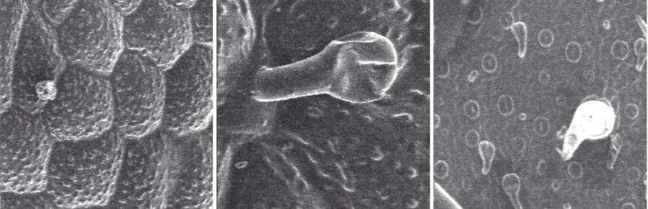
- View our live video of a green leaf inside the high vacuum system of the FE-SEM. Part I of the video shows the top side of the leaf with the cell walls and surface features clearly displayed (Figure 1). Note the partially deflated epidermal hairs on the top side of the leaf, located at the cell wall junctions (Figure 2).
- The second part of the video shows the bottom side of the leaf. Most noticeable are the circular features called stomata which regulate exchange of gasses (CO2, O2 and H2O) in and out of the cell (Figure 3). The number of stomata per square mm is characteristic of the plant species. We can quantitatively measure these in the SEM.
|